
Home/Dry Eye Prognosis and Complications
Dry Eye Prognosis and Complications
Learn about complications related to untreated dry eyes with TheraLife. Our clinically-proven, doctor-recommended formula works for 80% of first-time users.
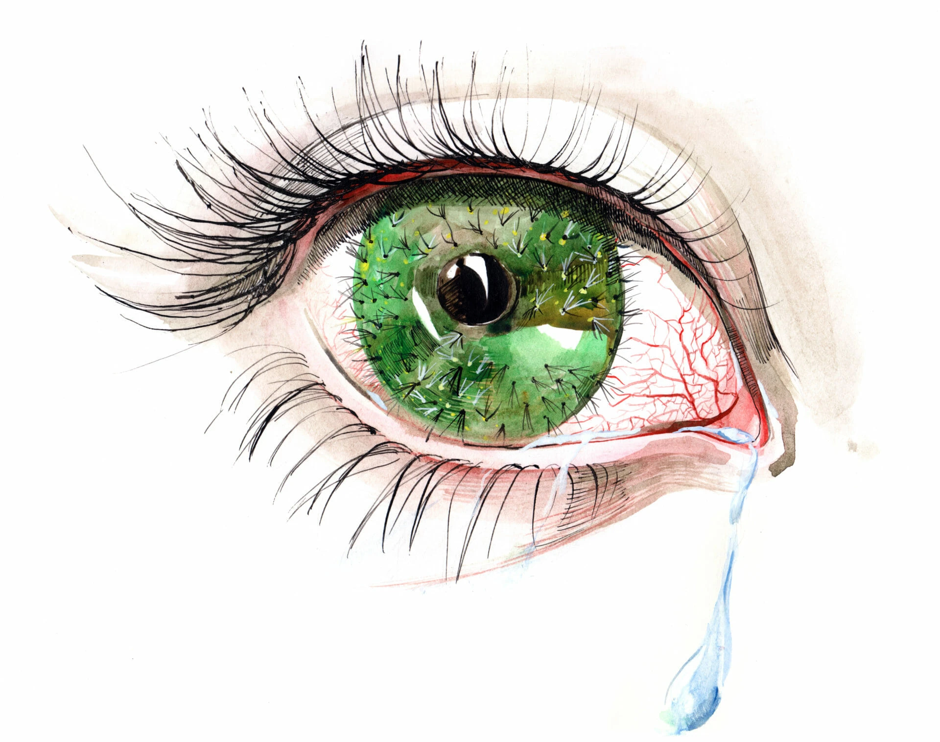
Most people don’t experience complications caused by dry eyes. If left untreated, however, dry eye syndrome can lead to serious complications. Complications that can occur include:
- More frequent eye infections. Your tears protect the surface of your eye from infection. Without adequate tears, you may have an increased risk.
- Scarring on the surface of your eye, in some cases requiring cornea transplant. Having dry eyes for a while can lead to tiny abrasions the surface of the eyes. In advanced cases, it can lead to loss of goblet cells. Some severe cases result in thickening of the corneal surface, corneal erosion, epithelial defects, corneal ulcerations (sterile and infected), corneal neovascularization, corneal scarring, corneal thinning, and even corneal perforation.
Dry eye syndrome usually is a chronic problem, especially if it is related to blepharitis or meibomian gland dysfunction. The prognosis for dry eyes shows considerable variance, depending upon the severity of the condition. Most people have mild-to-moderate cases and can be treated symptomatically with lubricants or oral capsules like TheraLife® Eye. This course of treatment provides a sustainable relief of symptoms and prevents future damages such as cornea aberrations, which could require cornea transplantation.
When dry eyes symptoms are severe, they can interfere with quality of life and cause depression. People sometimes feel their vision blurs with use, or they experience severe irritation to the point that they have trouble keeping their eyes open and may not be able to work or drive. Because of the debilitating impact they can have on daily functioning, dry eyes are now recognized as a common cause of depression.
Best Oral Dry Eye Treatment That Works When Drops Don't.

REFERENCES
- Stapleton F, Alves M, Bunya VY, et al. TFOS DEWS II epidemiology report. Ocul Surf 2017;15:334–65.
- Rolando M, Iester M, Macrì A, et al. Low spatial-contrast sensitivity in dry eyes. Cornea 1998;17:376–9.
- Benitez-Del-Castillo J, Labetoulle M, Baudouin C, et al. Visual acuity and quality of life in dry eye disease: proceedings of the OCEAN group meeting. Ocul Surf 2017;15:169–78.
- Uchino M, Schaumberg DA. Dry eye disease: impact on quality of life and vision. Curr Ophthalmol Rep 2013;1:51–7.
- Wolffsohn JS, Arita R, Chalmers R, et al. TFOS DEWS II diagnostic methodology report. Ocul Surf 2017;15:539–74.
- van Setten G, Labetoulle M, Baudouin C, et al. Evidence of seasonality and effects of psychrometry in dry eye disease. Acta Ophthalmol 2016;94:499–506.
- Nichols KK, Redfern RL, Jacob JT, et al. The TFOS international workshop on contact lens discomfort: report of the definition and classification subcommittee. Invest Ophthalmol Vis Sci 2013;54:14–19.
- Carnt NA, Keay L, Naduvilath T, et al. Risk factors associated with corneal inflammation in soft contact lens daily wear. Invest Ophthalmol Vis Sci 2007;49:4326.
- Rolando M, Cantera E, Mencucci R, et al. The correct diagnosis and therapeutic management of tear dysfunction: recommendations of the P.I.C.A.S.S.O board. Int Ophthalmol 2018;38:875–95.
- Baudouin C, Messmer EM, Aragona P, et al. Revisiting the vicious circle of dry eye disease: a focus on the pathophysiology of meibomian gland dysfunction. Br J Ophthalmol 2016;100:300–6.
- Mantelli F, Argueso P. Functions of ocular surface mucins in health and disease. Curr Opin Allergy Clin Immunol 2008;8:477–83.
- Craig JP, Tomlinson A. Importance of the lipid layer in human tear film stability and evaporation. Optom Vis Sci 1997;74:8–13.
- Rolando M, Refojo MF, Kenyon KR. Tear water evaporation and eye surface diseases. Ophthalmologica 1985;190:147–9.
- Braun RJ, King-Smith PE, Begley CG, et al. Dynamics and function of the tear film in relation to the blink cycle. Prog Retin Eye Res 2015;45:132–64.
- Stepp MA, Tadvalkar G, Hakt R, et al. Corneal epithelial cells function as surrogate Schwann cells for their sensory nerves. Glia 2017;65:851–63.
- Aragona P, Rania L, Puzzolo D. The role of inflammation, inflammatory cytochines, and ocular surface markers in dry eye disease. In: Benitez Del Castillo JM, Lemp MA, eds. Ocular surface disorders. London: JP Medical Publishing, 2013:55–68.
- Lemp MA, Baudouin C, Baum J, et al. The definition and classification of dry eye disease: report of the definition and classification subcommittee of the international dry eye workshop. Ocul Surf 2007;5:75–92.
- Musumeci M, Bellin M, Maltese A, et al. Chitinase levels in the tears of subjects with ocular allergies. Cornea 2008;27:168–73.
- Musumeci M, Aragona P, Bellin M, et al. Acidic mammalian chitinase in dry eye conditions. Cornea 2009;28:667–72.
- Chen Z, Tong L, Li Z, et al. Hyperosmolarity-induced codification of human corneal epithelial cells is regulated by JNK MAPK. Invest Ophthalmol Vis Sci 2008;49:539–49.